For an introduction to using NMR to analyze the structure of the peptide, start here.
Amino acid sequence of the peptide:
K1 T2 L3 T4 L5 E6 A7 A8 L9 R10 N11 A12 W13 L14 R15 E16 V17 G18 L19 K20
NMR spectra:
a) One-dimensional NMR spectrum of the peptide (500 MHz)
b) 2-D TOCSY spectrum.
Each cross peak indicates a pair of 1H nuclei located within the same amino acids. In general, strong peaks indicate 1H nuclei separated by 2 or 3 bonds; weaker peaks indicate 1H nuclei separated by > 3 bonds.
full_view tocsy
tocsy_section1.pdf
tocsy_section2.pdf
tocsy_section3.pdf
tocsy_section4.pdf
tocsy_section5.pdf
tocsy_section6.pdf
c) 2-D NOE spectrum, 80 msec mixing time.
In the 80 msec mixing time spectrum, each cross peak indicates a pair of 1H nuclei separated by less than about 4.5 angstroms, and not necessarily located within the same amino acid. Strong cross peaks indicate 1H nuclei separated by < 3 angstroms; weak cross peaks indicate pairs of protons separated by approximately < 4.5 angstroms.
full view noesy
noe section1.pdf
noe section2.pdf
noe section3.pdf
noe section4.pdf
noe section5.pdf
noe section6.pdf
noe section7.pdf
noe section8.pdf
noe section9.pdf
d) 2-D NOE spectrum, 400 msec mixing time.
In the 400 msec mixing time spectrum, the NOE effect extends to a longer range, by means of spin-diffusion. Strong cross peaks indicate 1H nuclei separated by approximately < 3.5 Å; weak cross peaks indicate pairs of protons separated by approximately < 6 Å.
noe (400ms) section1.pdf
noe (400ms) section2.pdf
noe (400ms) section3.pdf
noe (400ms) section4.pdf
noe (400ms) section5.pdf
noe (400ms) section6.pdf
noe (400ms) section7.pdf
noe (400ms) section8.pdf
noe (400ms) section9.pdf
e) 2-D COSY spectrum.
Each cross peak indicates a pair of 1H nuclei located within the same amino acids separated by 2 or 3 bonds.
cosy section1.pdf
cosy section2.pdf
cosy section3.pdf
cosy section4.pdf
cosy section5.pdf
f) A "PDB" coordinate file for the peptide is provided here. These coordinates are essentially a "random coil" starting structure, and can be refined using the information provided in the NMR spectra and X-PLOR (or other programs) to determine the actual structure of the peptide.
*************************************************
2. Useful things for the peptide NMR problem
Typical proton NMR chemical shifts for amino acids
Typical NMR chemical shifts for proteins:
H-1, C-13 & N-15 nuclei
Identifying amino acids in protein NMR spectra
Distances and torsion angles in regular
secondary structures
More distances in regular secondary structure
Typical NOE cross peaks in a helix
Typical NOE cross peaks in a beta strand
Typical NOE cross peaks in a beta sheet
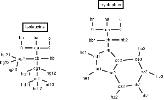
Naming of atoms in isoleucine and tryptophan
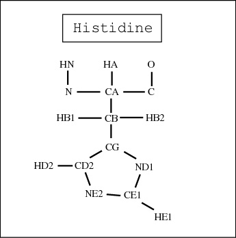
Naming of atoms in histidine
Using "Sparky" to evaluate volumes of
peaks in 2-D spectra
*****************************************************